Why Do Scleral Lenses Sometimes Get Foggy?
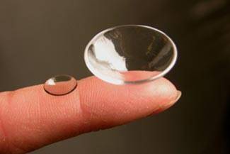
Scleral lenses provide a successful contact lens option for people with corneal conditions such as keratoconus, severe dry eye, and those needing vision correction following eye surgery. Unfortunately, about 30% of patients with scleral lenses may experience lens fogging. This requires them to remove their lenses and refresh the saline solution, that keeps their lenses lubricated, multiple times a day. Here are some reasons why scleral lenses fog up and tips on how to keep your lenses clear.
What is Midday Fogging?
Midday fogging is when scleral lenses fog up after a few hours of wear. The most likely causes appear to be an accumulation of debris from the tears between the lens and the cornea or an inflammatory reaction of the eye or eyelids to the contact lenses.
Fogging Caused by Debris
Blinking can sometimes cause the debris to dissipate, but it doesn’t always help. There are three types of tear debris that may lodge between the eye and the lens and cause fogging.
Mucin Debris
Mucin is an opaque, white, fluffy, oil-like layer of the tears. If the fit of the scleral lens isn’t perfect, mucin debris can move from the tears into the tear reservoir behind the scleral lens. If this is the case, your eye doctor will evaluate how the scleral lens fits and make the required adjustments to its design, most likely changing the peripheral edge lift.
The peripheral edge lift, the very edge of the scleral lens, allows a refreshing flow of tears to get under the lens and into the tear reservoir behind the scleral lens. However, if there is too much lift, excessive tears will flow, allowing debris to accumulate in the tear reservoir.
If the peripheral edge lift is the problem, the lens edge may be irritating your eyelid. Your eye doctor may ask you to reduce the amount of time you wear the lens, or have you remove and reapply the lens during the day. Another option is following a lens cleaning regimen using an enzymatic cleaner or a sodium hypochlorite-potassium bromide-based system.
Meibomian Debris
Meibomian glands are tiny glands in your eyelids that produce the essential oils for our eyes. Meibomian debris can be caused by Meibomian Gland Dysfunction (MGD) or blepharitis. The debris occurs when the oils of your tears find their way under the lens and appear as semi-transparent droplets of oil floating on water. It can disperse light, like an oil slick, or appear yellowish in color.
To reduce this form of debris your eye doctor will treat the underlying cause in the eyelids as well as review the lens design. If issues with meibomian debris persist, removing and reapplying the lens can help as well.
These types of debris can occur in combination, resulting in multiple management strategies.
Front Surface Debris
Front surface debris is any debris found on the outside of the scleral lens, from the buildup of protein to the debris mentioned above. External sources such as oil-based lotions, makeup, and face and hand soaps can also cause foggy vision. Knowing where the debris is coming from can help you and your eye doctor eliminate the problem.
To remove foggy vision, make sure to wash your hands with mild hand soaps, and then rinse before handling your lenses. Also, make sure to apply face cream or makeup after inserting your lenses. Avoiding oil-based moisturizers on the eyelids, and not applying makeup to the inside area of the eyelid margin or over the meibomian glands can decrease the risk of MGD or obstruction.
Fogging Due to Inflammation
Giant Papillary Conjunctivitis (GPC)
GPC is an inflammatory reaction caused by the contact lenses. It occurs in up to 15% of all hard lens wearers and is likely due to the edge of the lens rubbing up against the conjunctiva, the protective layer on the eyelids and the outside part of the eyes. The signs of GPC are red, swollen and irritated eyelids.
If you have GPC your eye doctor may alter the design of your lens, most commonly the peripheral edge lift, and prescribe mast cell stabilizer antihistamine drops or reduced lens wearing time. The doctor may also do a deep clean of the lens with a sodium hypochlorite-potassium bromide-based system with enzymatic cleaners.
Atopic Disease and Keratoconus
Another type of debris that someone might experience with scleral lenses is due to an association between atopic disease (typically associated with the immune response to common allergens). This type of debris appears as a diluted milk-like fog in the scleral lens fluid reservoir under the lens.
Your eye doctor may recommend the following treatment options, including: reducing excessive edge lift, reducing base curves, taking an antihistamine to reduce inflammation, lens removal and reapplication, or in extreme cases, topical steroids.
Scleral lenses can be a great option for many patients, even if fogging occurs. These management strategies, along with proper lens care, can go a long way to ensure healthy life-long scleral lens wear. Contact Kelly Vision Center to determine what may be causing your foggy vision and how to treat it today!
Our practice serves patients from Goodlettsville, Hendersonville, Gallatin, and Madison, Tennessee and surrounding communities.


